Brain Routine (CT) PRINT
Updated: 8/16/2021
What Changed: Request for bone kernel axial at 1-1.25 mm rather than 2-3 mm given the amount of trauma seen at hospitals and need to evaluate for non-displaced fractures.
History: Alteration of consciousness, altered mental status, headache, seizure, mass, trauma, intracranial hemorrhage, stroke, hydrocephalus, herniation.
| BRAIN ROUTINE | ||
|---|---|---|
| Localizer | Frontal and Lateral | |
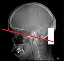
| Angulation | Supraorbito-meatal line | |
| Scan type | axial / sequential (GE + Siemens), helical (Toshiba + Cannon) | |
| Target CTDIvol (mGy) | <45 | |
| Max CTDIvol (mGy) | <70 | |
| Max scan time | 30 sec | |
| Detector/Slice | Detector minimum | |
| kVp | 100-140 | |
| Pitch | N/A | |
| DFOV | 24.0-25.0 | |
| Coverage | ||
| Ax | foramen magnum to vertex | |
| Cor | nose to occiput | |
| Sag | ear to ear | |
| Contrast: very rare need for contrast (e.g. scalp or face soft tissue mass). have a low threshold to call radiologist to confirm need | ||
| Agent | 80 mL (350/ 370 concentrate ) or 100 mL (300 concentrate) | |
| Delay | 120 sec | |
| Rate | power inject 2-3 mL / sec OR hand inject 1-1.5 mL / sec | |
| SEND TO PACS | ||
| Ax 2-3 mm soft | ||
| Ax 1-1.25 mm bone | ||
| Cor and Sag 2-3 mm soft | ||
Key tips:
-
- For GE, Siemens, Phillips, helical scanning of the brain should be avoided unless it is the only option available for obtaining diagnostic information. Scan in standard 360 degree axial or contiguous fashion. However, for Canon and Toshiba scanners, helical scanning may be preferred for dose and image quality purposes.
-
- Scan with “Head” SFOV or comparable corresponding bowtie filter to correct for cupping artifact at skull / brain interface (can mimic a subdural hematoma).
-
- Image-guided surgery CT Scans (Brainlab, Stryker, stealth, InstaTrack). Follow slice thickness, spacing, coverage and FOV guidelines provided by individual vendors. Typically this involves the use of non-‐angled slices and meticulous patient positioning for scans with 0.6-1.25mm contiguous slices. The images are typically also provided to the referrer on a CD in an uncompressed DICOM format.
- Ideal angulation parallel to a line intersecting the upper 3rd of the orbit and the foramen magnum (right image below). A representative lowest slice avoids the lens (left image below). Avoid the eye lens if possible since it is sensitive to radiation. This is the expected angle of axials under ideal circumstances.
Coverage Examples:
Bottom Slice 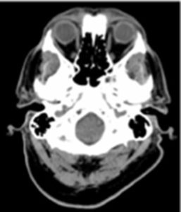
Axial angle