Cervical Spine (CT) PRINT
Last updated: 8/16/2021.
What changed: For outpatient exams, request to send 1 mm sag in soft tissue kernel to help radiologist evaluate degenerative change.
History: Pain, stenosis, trauma, infection, scoliosis, post-operative evaluation
| CERVICAL SPINE | ||
|---|---|---|
| Localizer | Frontal and Lateral | |
| Patient Instructions | Do not swallow | |
| Coverage | Foramen magnum through T1 vertebra | |
| Scan type | Helical | |
| kVp | 120-140 | |
| Target CTDIvol (mGy) | <25 | |
| Max CTDIvol (mGy) | 40 (no hardware); 50 (if hardware) | |
| Max scan time (sec) | 30 | |
| Pitch | 0.8-1.5:1 | |
| DFOV | 13-16 | |
| SEND TO PACS | ||
| Ax: 1-1.25 mm Contiguous Bone | ||
| Ax: 1-1.25 mm Contiguous Standard | ||
| Sag and Cor: 1 mm Bone | ||
| Sag: 1 mm soft tissue | ||
| If metallic hardware: VRT with semi-transparent bone & opaque metal | ||
- Contrast should not be needed in most cases to evaluate for trauma or degenerative changes. If ordered, call a radiologist to confirm.
- Myelogram: ask radiologist whether they want prone or supine images.
Key tips:
- Have the patient lower their shoulders for the cervical spine scans, even if using tension Velcro straps designed for this purpose. It both improves image quality and reduces dose.
- Only include one vertebral element above and below target.
- On axials, the target is the canal, not the anterior neck.
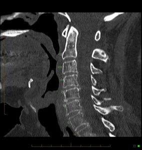
Coverage Examples:
Axial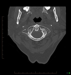
Coronal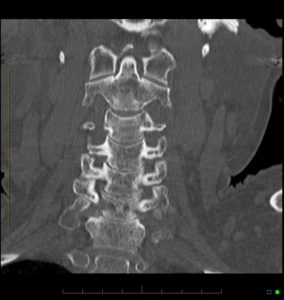
Sagittal