Temporal Bones (CT) PRINT
Version: 1.0
Last updated: 3/11/2021.
What changed: Updated Stenvers coverage example.
History: cholesteatoma, trauma, otomastoiditis, hearing loss, otosclerosis
Without Intravenous Contrast
| TEMPORAL BONES WITHOUT | ||
|---|---|---|
| Localizer | Frontal and lateral | |
| Angulation | orbitomeatal line pependicular to table; TMJ perpendicular to table | |
| Scan type | helical | |
| kVp | 120 | |
| Target CTDIvol (mGy) | <60 (adult); 40-55 (peds) | |
| Max CTDIvol (mGy) | 68 | |
| Max scan time | 35 sec | |
| Pitch | 0.9-1.5:1 | |
| DFOV | 17-18 (bilat); 10 (unilat) | |
| Coverage | mastoid tip to petrous ridge | |
| SEND TO PACS | ||
| Ax Bilateral: 0.5-0.625 mm Contiguous bone scanned in plane; DFOV 17-18 | ||
| Ax Targeted R and L: 0.5-0.625 mm with interval 0.4-0.5 mm and DFOV 10; in plane with lateral semicircular canal | ||
| Cor Targeted R and L: 0.5-0.625 mm with interval 0.4-0.5 mm and DFOV 10; perpendicular to axial | ||
| Ax Bilateral: 2 mm soft scanned in plane; DFOV 17-18 | ||
| Poschl & Stenvers if indication mentions dehiscence | ||
With Intravenous Contrast
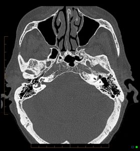
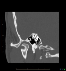
Coverage Examples:
Bilateral axial followed by unilateral axial and coronal images:
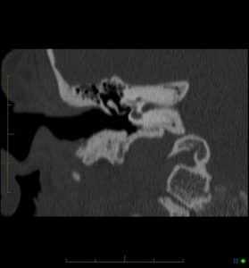
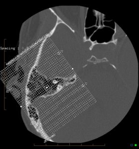
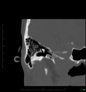
Poschl: Oblique sagittal setup / scout is aligned to superior semicircular canal.
Planning
You’ve done it correctly if you can see the entire superior semicircular canal in a single image plane
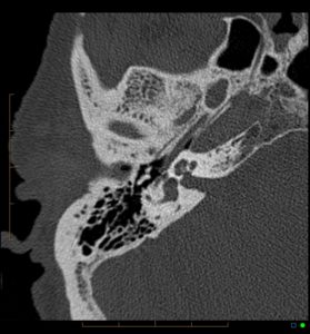
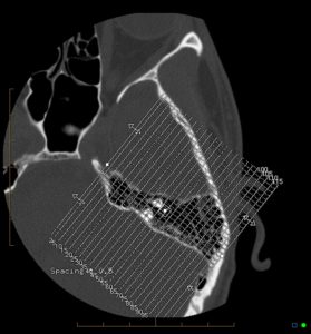
Stenvers : Oblique coronal setup is aligned perpendicular to Poschl plane. Series should cover the central portion of the temporal bone.
Planning