Thoracic Spine (CT) PRINT
Last updated: 8/16/2021.
What changed: send soft tissue axial AND sagittal at 2 mm thick.
History: Pain, degenerative change, trauma, post-operative evaluation, infection.
Contrast should not be needed in most cases to evaluate for trauma or degenerative changes. If ordered, call a radiologist to confirm.
Myelogram: ask radiologist whether they want prone or supine images.
Discogram field of view includes two vertebral levels above and below injected disc
Coverage Examples:
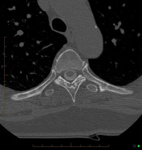
Axial: don't need much beyond canal
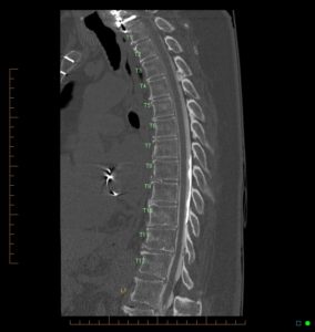
Sagittal: don't include more than C7 - L1